Go back
SWIp in traumatic brain injury
Patient information
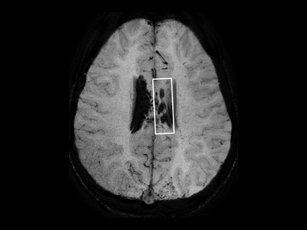
In this case, a 10-year-old girl thrown from a horse. The SWIp images provided increased visibility of the corpus callosum injury compared to the T2-weighted, diffusion weighted and gradient echo images, see the box in the images. SWIp also provides increased visibility of the cortical contusion (arrows) compared to gradient echo imaging. In this case, SWIp helped to characterize the extent of the patient’s injury, which is important to know for short term care and longer term prognosis and rehabilitation.Gallery
More Information
- Brochure: Clinical applications
- Video: MRI Neuro Highlights and Clinical Advancements at Dent Neurologic Institute
- FieldStrength article: Ingenia 3.0T delivers high performance MRI to the busy practice at DMG
- FieldStrength article: Recently adopted methods for neuro MR improve efficiency and confidence
- FieldStrength article: High quality imaging in MS, stroke and brain tumor
- FieldStrength article: UVM brain MRI protocols upgraded with latest methods
- Compressed SENSE in practice: MR in the Emergency Department
- Customer testimonial: Dr Tetsuya Yoneda, Kumamoto University, Japan - Collaboration on SWIp
- Customer testimonial: Dr Chip Truwit, Hennepin County Medical Center, Minneapolis, USA - SWIp integral part of all MR trauma scans
*Results from case studies are not predictive of results in other cases. Results in other cases may vary.


